Developing procedural techniques to visualize cell growth with accuracy and realistic beauty.
Our team of 3D wizards love a visualization challenge, and the biological processes at the core of many of our animations provide a fun and complex playground.
For Research into the biology and morphology, we lean on our scientific background while also gathering more detailed information from textbooks, publications, and histology images specific to each project.
For the 3D and motion Development, our primary 3D software tools are Cinema4D, X-particles, and a dash of Houdini. When we’re stuck, we look to 3D tutorials for inspiration.
Mitosis Tests
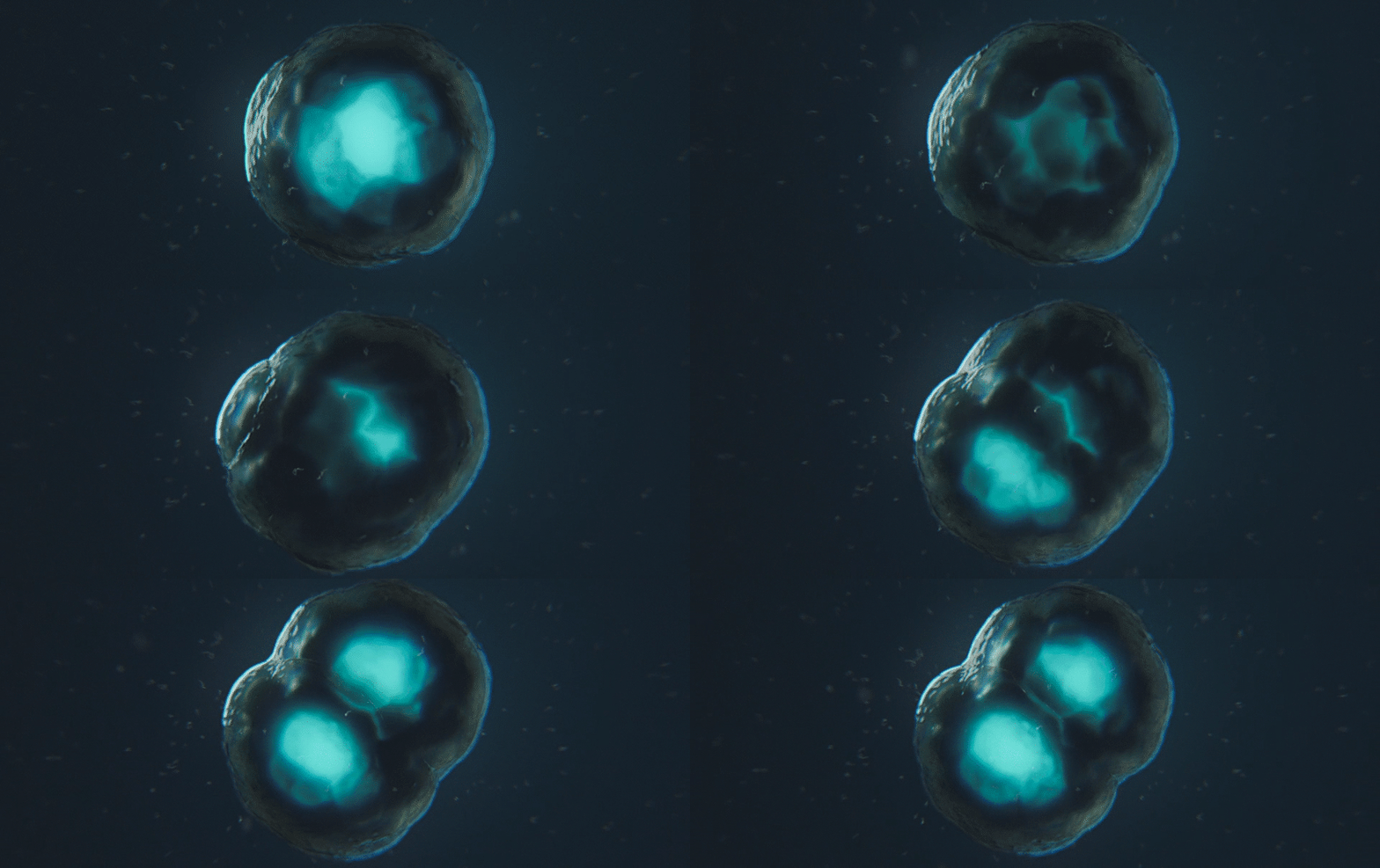
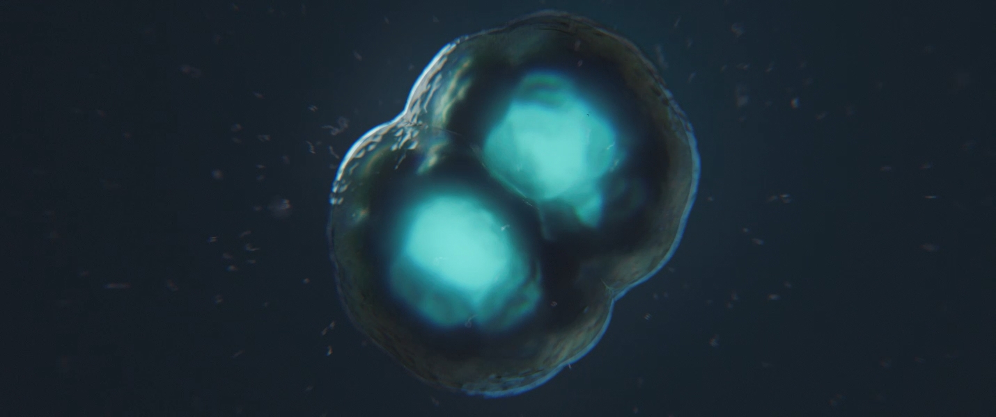
Previews of an embryonic development animation – the moment a zygote cleaves into two cells and begins the journey towards a multicellular organism.

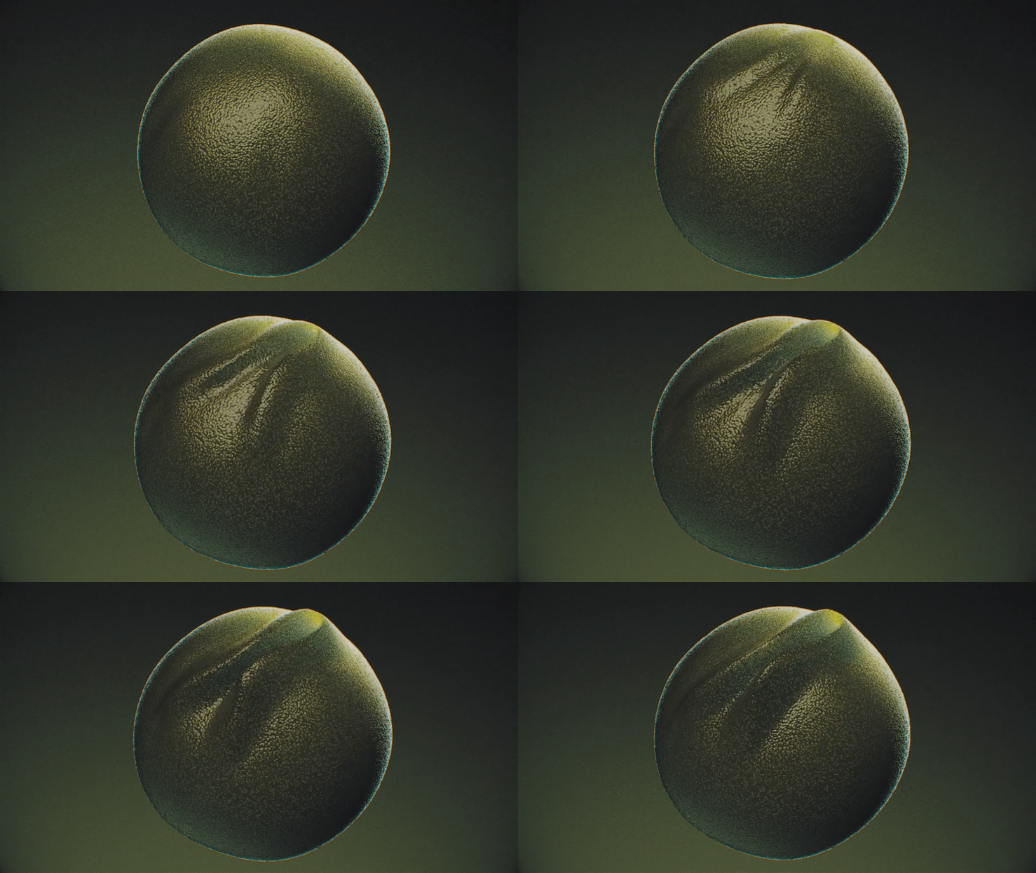
Neural crest development, prior to neural tube formation in this mysterious embryo.

Hyperplasia Tests
For a mechanism of disease project, we needed to depict the rapid division and proliferation – hyperplasia – of basal epithelial cells. Viral-inclusion bodies (the yellow spheres) also assemble and grow inside these particular cells over time.
More news

UIC Alumni Interview with Mike Moran

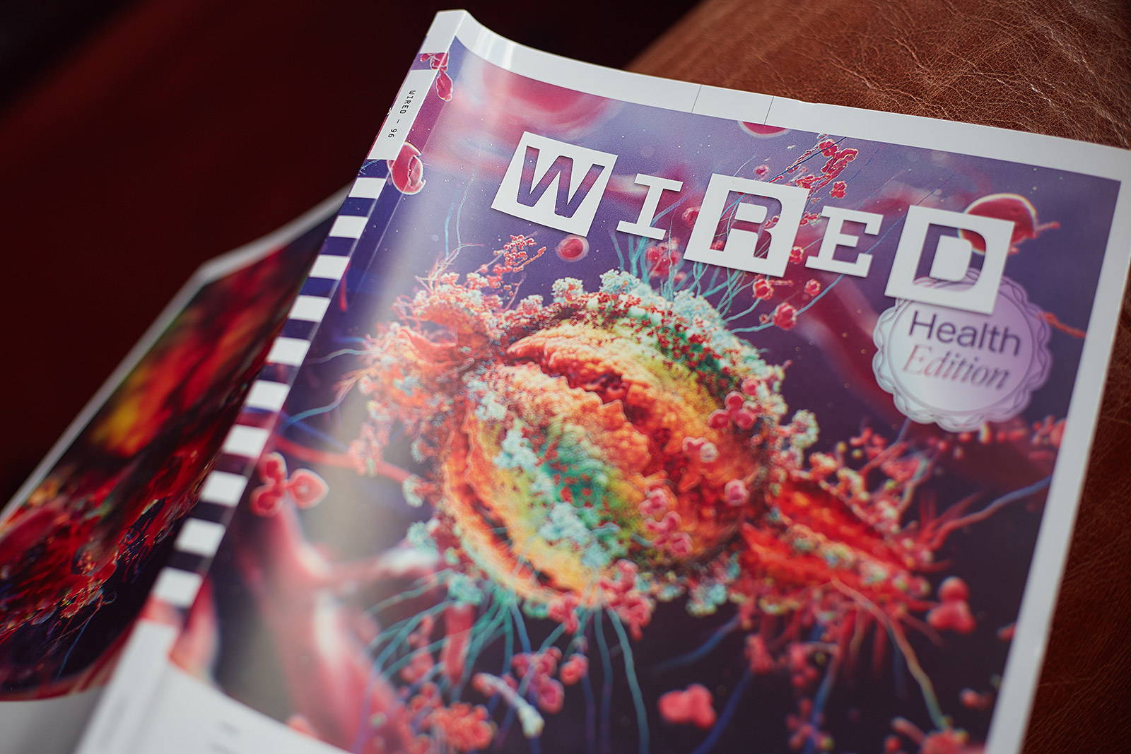
ON THE COVER OF WIRED MAGAZINE

Newt Studios Presents at ZBrush Summit 2019

